Nova Eye Medical

Lighting the way in canaloplasty

Learn more

Canaloplasty, designed around physiology

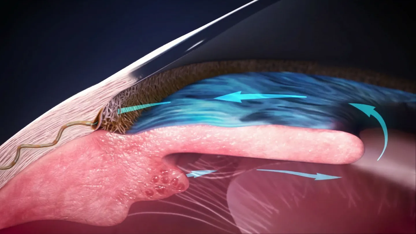
Canaloplasty is a minimally invasive glaucoma surgical procedure designed to improve conventional outflow using 360° microcatheterization and pressurized viscodilation. The goal is to enhance flow through the trabecular meshwork, Schlemm’s canal and collector channels while working with the eye’s natural drainage anatomy. Microcatheter-based systems such as iTrack™ Advance support this approach with illuminated navigation and pressurized viscoelastic delivery.
1. Grieshaber, M. C., Pienaar, A., Olivier, J., & Stegmann, R. (2010). Clinical evaluation of the aqueous outflow system in primary open-angle glaucoma for canaloplasty. Investigative Ophthalmology & Visual Science, 51(3), 1498–1504.Manik Goel, Renata GPicciani, Richard KLee, and Sanjoy KBhattacharya. Aqueous Humor Dynamics: AReview. Open Ophthalmol J. 2010; 4: 52 – 59.


2. Smit, B. A., & Johnstone, M. A. (2002). Effects of viscoelastic injection into Schlemm's canal in primate and human eyes: Potential relevance to viscocanalostomy. Ophthalmology, 109(4), 786–792.
Honeycomb green icon

Designed To Work With Natural Outflow Physiology

Canaloplasty addresses the conventional outflow pathway — viscodilating Schlemm's canal and opening collector channels to restore the eye's natural drainage system (1). No implant—just a procedure designed to treat the underlying resistance, not bypass it. (2)

Fingerprint green icon

Hardware-free

Dilates the canal without leaving hardware in the eye, leaving the door open for future interventions

institution green icon

Trusted for over two decades

A surgical legacy stretching back to 2004, with over 100,000 procedures performed worldwide

Canaloplasty infographic
Schlemm’s Canal
The trabecular meshwork and beyond
360° catheterization and viscodilation aim to expand the canal and address focal narrowing along its circumference.
2
Trabecular Meshwork
A key site of outflow resistance
Canaloplasty is designed to gently separate compressed trabecular structures to support aqueous passage.
1
Collector Channels
Three points of outflow resistance
Pressurized viscoelastic delivery is intended to promote broader distribution toward collector channel ostia, supporting flow beyond Schlemm’s canal.
3
Honeycomb green icon

Designed To Work With Natural Outflow Physiology

Canaloplasty addresses the conventional outflow pathway — viscodilating Schlemm's canal and opening collector channels to restore the eye's natural drainage system (1). No implant—just a procedure designed to treat the underlying resistance, not bypass it. (2)

Fingerprint green icon

Hardware-free

Dilates the canal without leaving hardware in the eye, leaving the door open for future interventions

institution green icon

Trusted for over two decades

A surgical legacy stretching back to 2004, with over 100,000 procedures performed worldwide

1. Grieshaber, M. C., Pienaar, A., Olivier, J., & Stegmann, R. (2010). Clinical evaluation of the aqueous outflow system in primary open-angle glaucoma for canaloplasty. Investigative ophthalmology & visual science, 51(3), 1498–1504.

2. Smit, B. A., & Johnstone, M. A. (2002). Effects of viscoelastic injection into Schlemm's canal in primate and human eyes: potential relevance to viscocanalostomy. Ophthalmology, 109(4), 786–792.

What does canaloplasty do differently to other glaucoma procedures?

Rather than attempt to mechanically change or bypass the pathway of aqueous outflow, canaloplasty acts tore-establish the natural flow of aqueous throughout 360° of the conventional outflow pathway. Akin to angioplasty of the eye, iTrack™ combines aprocess of catheterization and pressurized viscodilation to address all potential points of blockage in the trabecular meshwork, Schlemm’s canal and the collector channels.
Quote Gradient Icon
Canaloplasty is the best example of early glaucoma intervention.
Larissa Camejo, MD (USA)
Cataract surgery, glaucoma management
Center for Medical and Surgical Eye Care, Palm Beach Gardens, Florida
Dr Larissa Camejo BW photo
Quote Gradient Icon
Canaloplasty comprehensively treats outflow locations, which is why it is my first go-to glaucoma procedure. I don't have the diagnostic capability to know where the obstruction is located or what level of resistance exists, and so I like to start off with a glaucoma procedures that addresses everything.
Mahmoud A. Khaimi, MD
Glaucoma Specialist
Dean McGee Eye institute, University of Oklahoma
Mahmoud Khaimi BW photo

How is canaloplasty different from other glaucoma procedures?

Compare canaloplasty with other glaucoma approaches across implants, tissue interaction and outflow pathway coverage.
iTrack Advance™ Logo Black
Other glaucoma procedures
Implant-free (no permanent device left behind)
Gradient Check Icon
Unavailable symbol Grey Icon
Tissue-preserving
Gradient Check Icon
Unavailable symbol Grey Icon
Targets all three resistance sites (trabecular meshwork, Schlemm's canal, and collector channels)
Gradient Check Icon
Unavailable symbol Grey Icon
Physiological restoration of natural aqueous outflow
Gradient Check Icon
Unavailable symbol Grey Icon

1. Glaucoma Specialists & iTrack. "Discover iTRACK™ for Glaucoma." https://glaucoma-itrack.com/us/.
2. Glaucoma Associates of Texas. "Gonioscopy-Assisted Transluminal Trabeculotomy (GATT)." https://www.glaucomaassociates.com/gonioscopy-assisted-transluminal-trabeculotomy/.
3. Lewis, R.A., et al. "Canaloplasty: Three-Year Results." Journal of Cataract & Refractive Surgery, vol. 37, no. 4, 2011, pp. 682-690. PMID: 21420593. https://pubmed.ncbi.nlm.nih.gov/21420593/

Targets multiple resistance sites: Canaloplasty addresses all three sites: trabecular meshwork, Schlemm's canal, and collector channels. Most other MIGS address only 1-2 sites; stents primarily address trabecular meshwork, while TM-cutting procedures address only trabecular meshwork.
1.Banerjee, A., et al. "Comparison of Superior versus Inferior Canaloplasty and Goniotomy with OMNI Surgical System." PLoS ONE, 2024. https://pmc.ncbi.nlm.nih.gov/articles/PMC11214751/
2. Patel, K., & Reiss, G. "Long-Term Clinical and Safety Outcomes of Canaloplasty Performed with iTrack Microcatheter." PLoS ONE, 2023. https://pmc.ncbi.nlm.nih.gov/articles/PMC10191748/
3. Meta-analysis authors. "Safety and Efficacy of Canaloplasty versus Trabeculectomy in Open-Angle Glaucoma." Oncotarget, 2019. https://www.oncotarget.com/article/14757/text/

Physiological restoration of natural aqueous outflow: Canaloplasty rejuvenates and re-establishes the eye’s natural drainage system across the entire conventional outflow pathway without creating artificial bypasses, shunts, or tissue removal.
1. Arosemena A, Simpson RG, Bedrood S, Camejo L, Funke CM, Kamat S, Qiu M. Canaloplasty: how it works and where it fits in the MIGS armamentarium. Glaucoma Today. 2025 Mar/Apr suppl. https://glaucomatoday.com/articles/2025-mar-apr-supplement/canaloplasty-how-it-works-and-where-it-fits-in-the-migs-armamentarium

More on canaloplasty

Canaloplasty experts share a quick word on what canaloplasty means to them.
Drs. Ike Ahmed, Paul Singh and more talk about canaloplasty in the clinic.

What the experts say about canaloplasty with iTrack™ Advance

Nova Eye Medical, equips your surgeons with the best equipment for a non-destructive, long-term, patient care focused solution.


Quote symbol in white
Canaloplasty is the best example of early glaucoma intervention.
Larissa Camejo
Senior Glaucoma Specialist
Quote symbol in white
If you want to perform a minimally invasive procedure without causing any form of damage, and without leaving any foreign body in the eye, canaloplasty is the best option.
Karl Mercieca
Quote symbol in white
The collector channels go 360 degrees around, and I think that's one of the huge advantages of iTrack™ Advance and canaloplasty—being able to treat 360º in one fluid treatment
Morgan Micheletti
MD, FACS
Quote symbol in white
That’s why canaloplasty makes so much sense: We’re treating the problem tissue in the eye, and more to the point, because of the high degree of safety, we are doing so earlier in the disease continuum.
Robert Noecker
MD, MBA
Arrow pointing Left white icon
White Arrow Pointing Right

Contact us

Not all products are available in all regions or countries. Please contact your Nova Eye representative for more information.
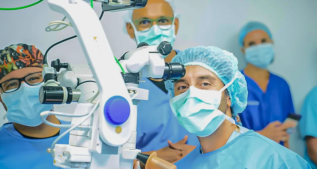
Surgeons in the Operating room using itrack devices
Thank you! Your submission has been received!
Oops! Something went wrong while submitting the form.

Disclaimer

North America

The iTrack™ Advance has a CE Mark (Conformité Européenne) 2862 and US Food and Drug Administration (FDA) 510(k) #K221872 for the treatment of open-angle glaucoma.

INDICATIONS (USA): The iTrack™ Advance is indicated for fluid infusion or aspiration during surgery. The Nova Eye iTrack™ Advance is indicated for the cutting or disruption of the trabecular meshwork during goniotomy procedures.* The Nova Eye iTrack™ Advance is indicated for catheterization and viscodilation of Schlemm’s canal to reduce intraocular pressure in adult patients with open-angle glaucoma.The ab interno surgical technique is not a cleared indication of the iTrack™ Advance in the USA.

*The iTrack™ Advance cutting function (goniotomy) is a Class 1 510(k) exempt device function that is not specifically indicated for the reduction of intraocular pressure (IOP) or the treatment of open-angle glaucoma.

CONTRAINDICATIONS (USA): The iTrack™ Advance is not intended to be used for catheterization and viscodilation of Schlemm’s canal to reduce intraocular pressure in eyes of patients with the following conditions: Neovascular glaucoma; Angle-closure glaucoma; Previous surgery with resultant scarring of Schlemm’s canal.

PRECAUTIONS (USA): The iTrack™ Advance should be used only by physicians trained in ophthalmic surgery. Knowledge of surgical techniques, proper use of the surgical instruments, and post-operative patient management are considerations essential to a successful outcome.


© 2026. Nova Eye, Inc. E&OE. Patents pending and/or granted. iTrack™ Advance, iTrack™, ViscoInjector™ and iLumin™ are trademarks of Nova Eye, Inc.

Disclaimer

North America

The iTrack™ has a CE Mark (Conformité Européenne) 2862 and US Food and Drug Administration (FDA) 510(k) #K080067 for the treatment of open-angle glaucoma.

INDICATIONS: The iTrack™ canaloplasty microcatheter is indicated for fluid infusion or aspiration during surgery. The iTrack™ canaloplasty microcatheter is indicated for catheterization and viscodilation of Schlemm’s canal to reduce intraocular pressure in adult patients with open-angle glaucoma.The ab interno surgical technique is not a cleared indication for the iTrack™ in the USA.

CONTRAINDICATIONS: The iTrack™ canaloplasty microcatheter is not intended to be used for catheterization and viscodilation of Schlemm’s canal to reduce intraocular pressure in eyes of patients with the following conditions: neovascular glaucoma; angle closure glaucoma; and, previous surgery with resultant scarring of Schlemm’s canal.

ADVERSE EVENTS: Possible adverse events with the use of the iTrack™ canaloplasty microcatheter include, but are not limited to: hyphema, elevated IOP, Descemet’s membrane detachment, shallow or flat anterior chamber, hypotony, trabecular meshwork rupture, choroidal effusion, Peripheral Anterior Synechiae (PAS) and iris prolapse.

WARNINGS: The iTrack™ canaloplasty microcatheter is intended for one time use only. DO NOT re-sterilize and/or reuse, as this can compromise device performance and increase the risk of cross contamination due to inappropriate reprocessing.

PRECAUTIONS: The iTrack™ canaloplasty microcatheter should be used only by physicians trained in ophthalmic surgery. Knowledge of surgical techniques, proper use of the surgical instruments, and post-operative patient management are considerations essential to a successful outcome.
Get the latest Nova Eye Medical news alerts, straight to your inbox

Thank you for subscribing to our newsletter!
Oops! Something went wrong while submitting the form. Please try again.

Get the latest Nova Eye Medical news alerts, straight to your inbox

Thank you for subscribing to our newsletter!
Oops! Something went wrong while submitting the form. Please try again.
Scroll to the top white icon