Clinical Applications

This page provides a summary of the iTrack™ Advance clinical applications by region. Indications and available device functions vary depending on where the device is used. For complete information, including contraindications, warnings, precautions and adverse events, please refer to the Instructions for Use (IFU).

The iTrack™ Advance has a CE Mark (Conformité Européenne) for the treatment of open-angle glaucoma.

Indications for use

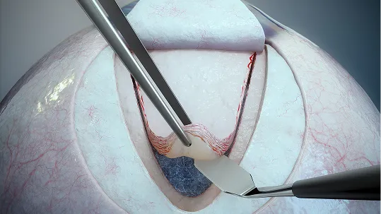
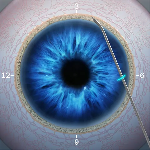
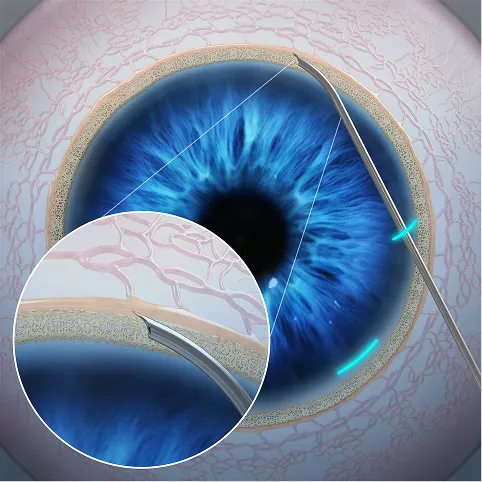
The iTrack™ Advance is indicated for fluid infusion or aspiration during surgery. The iTrack™ Advance is indicated for catheterization and viscodilation of Schlemm’s canal to reduce intraocular pressure in adult patients with open-angle glaucoma.

About Canaloplasty

Canaloplasty is a microcatheter-based glaucoma procedure that uses 360° catheterization of Schlemm’s canal followed by viscodilation with viscoelastic. The goal is to support aqueous outflow through the conventional pathway by addressing resistance along the trabecular meshwork, Schlemm’s canal and collector channels.

Ab-Interno Canaloplasty

About the ab-interno approach

In an ab-interno approach, Schlemm’s canal is accessed from within the anterior chamber under gonioscopic visualization. The procedure is performed through a small corneal incision, enabling canal catheterization and viscodilation while avoiding external conjunctival dissection.

Ab-interno canaloplasty with iTrack™ Advance

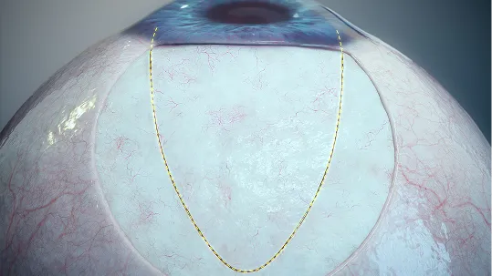
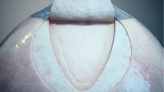
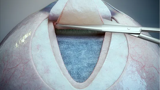
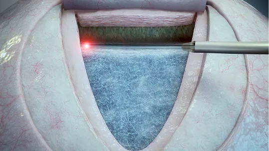
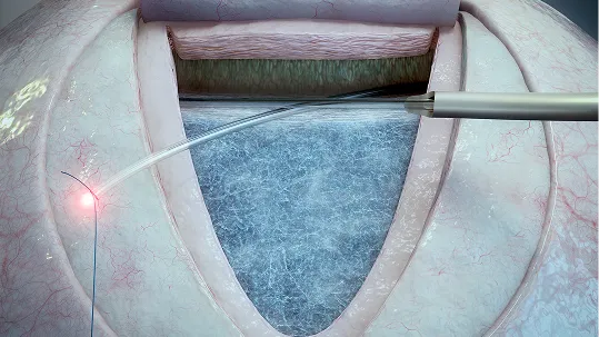
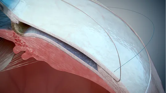
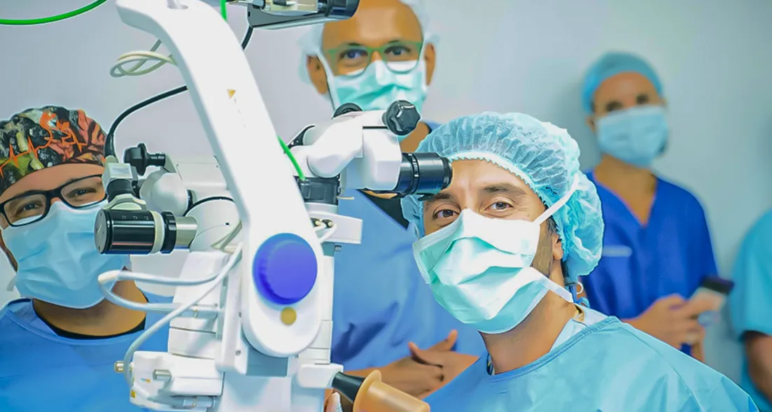
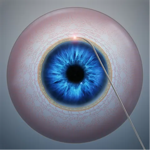
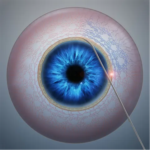
With iTrack™ Advance, ab-interno canaloplasty is performed by accessing Schlemm’s canal from within the eye, advancing a microcatheter through the canal and delivering viscoelastic to viscodilate the pathway as the catheter is withdrawn. The steps below summarize the procedural concept at a high level. For full details, refer to the IFU.

Step 1
Corneal incision
Under gonioscopic visualization, Schlemm’s canal is accessed through a small corneal incision and the microcatheter is introduced into the canal.
Ab interno Step 1
Step 2
Cannula insertion
Using the cannula tip, pierce the upper most anterior portion of pigemented trabecular meshwork at a 15° angle. Release the forward pressure with the cannula tip. Rest the cannula tip against the scleral wall. Take care to remain as static as possible. Ensure the cannula is orientated parallel with Schlemm’s canal.
Ab interno Step 2
Step 3
Canal intubation
Use the actuator to slowly advance microcatheter into Schlemm’s canal. If resistance is felt, re-adjust the position of the cannula tip and gently try again. Verify microcatheter is in Schlemm’s canal via the illuminated tip.
Ab interno Step 3
Step 4
Cannula insertion
Using the cannula tip, pierce the upper most anterior portion of pigemented trabecular meshwork at a 15° angle. Release the forward pressure with the cannula tip. Rest the cannula tip against the scleral wall. Take care to remain as static as possible. Ensure the cannula is orientated parallel with Schlemm’s canal.
Ab interno Step 4
Step 5
Viscodilation
Use actuator to slowly withdraw microcatheter from Schlemm’s canal while instructing scrub nurse to simultaneously deliver viscoelastic via the ViscoInjector.
Healon Pro: 9 clicks per quadrant (36 clicks over 360°)ProVisc: 9 clicks per quadrant (36 clicks over 360°)
Ab interno Step 5
Download ab-interno PDF
Download white icon
Download information
Download white icon

Ab-Externo Canaloplasty

About the ab-externo approach

In an ab-externo canaloplasty technique, Schlemm’s canal is accessed through a scleral approach. The canal can then be intubated with a microcatheter to support catheterization and viscodilation of Schlemm’s canal as part of a canaloplasty procedure.

Ab-externo canaloplasty with iTrack™ Advance

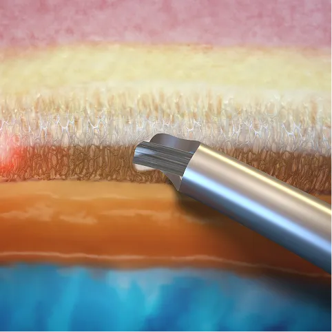
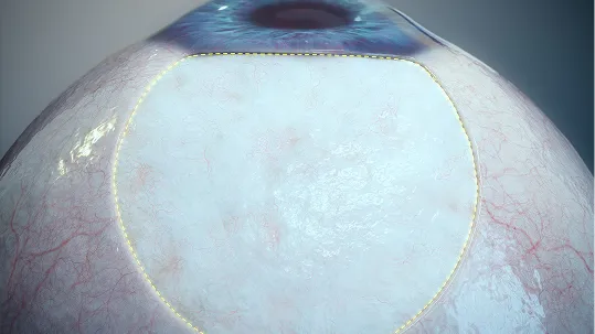
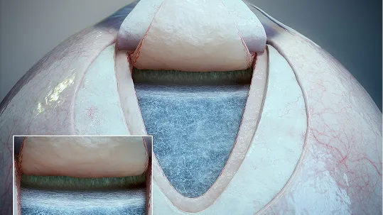
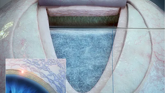
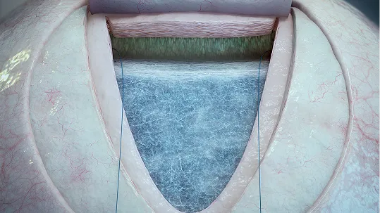
Most glaucoma treatments fail to completely address the natural outflow system and may even disturb the natural outflow function. Unlike traditional glaucoma surgeries (trabeculectomy and tube shunts), which bypass the natural outflow system, canaloplasty works by restoring the natural ocular outflow function in four keysteps:

Step 1
Creation of the outer flap to enable access for the ab-externo approach.
Ab Externo Procedure imageAb Externo Procedure imageAb Externo Procedure image
Step 2
Creation of the inner flap to expose Schlemm’s canal and prepare for intubation.
Ab Externo Procedure imageAb Externo Procedure imageAb Externo Procedure image
Step 3
Intubation of Schlemm’s canal with the microcatheter, with catheterization and viscodilation performed per the IFU.
Ab Externo Procedure image
Step 4
Suture placement, followed by outer flap closure using standard technique.
Ab Externo Procedure imageAb Externo Procedure imageAb Externo Procedure image
Step 5
The outer flap is closed to ensure precise alignment and healing.
Ab Externo Procedure imageAb Externo Procedure image
Download ab-externo PDF
Download white icon
Download information
Download white icon

Disclaimer

Contact us

Not all products are available in all regions or countries. Please contact your Nova Eye representative for more information.
Surgeons in the Operating room using itrack devices
Thank you! Your submission has been received!
Oops! Something went wrong while submitting the form.

Contact us

Thank you! Your submission has been received!
Oops! Something went wrong while submitting the form.
*In-house bench testing data using a proprietary optical rheology test apparatus to assess the change in viscosity over time during viscoelastic delivery with the iTrack™ Advance. Bench testing data may not be indicative of clinical outcomes. Clinical effects depend on anatomy and surgical technique. Results may vary.